Posterior Rib Cage Muscles / Thoracic Muscles Attachments Actions Teachmeanatomy - Rectus capitis posterior major, rectus capitis posterior minor, obliquus capitis superior, obliquus capitis inferior.
byAdmin•
0
Posterior Rib Cage Muscles / Thoracic Muscles Attachments Actions Teachmeanatomy - Rectus capitis posterior major, rectus capitis posterior minor, obliquus capitis superior, obliquus capitis inferior.. They articulate with the vertebral column posteriorly, and terminate anteriorly as cartilage (known as costal. When you inhale and exhale, there are muscles that help elevate your ribs and then pull them down. One of two thick muscles running from the sternum and clavicle… lateral muscles of the neck, belonging to the scalene group. A large left pneumothorax is present (arrows). Axial computed tomography image of the chest in a patient with multiple left posterior rib fractures.
Rib cage muscles (page 1). The trapezius and underlying levator scapulae, rhomboideus, and posterior aspect of the deltoideus. Serratus anterior, serratus posterior superior, serratus posterior inferior. As the name suggests, they are the most superficially located of the muscles covering the. The serratus rotates the inferior angle of the scapulae, protracts the scapulae laterally toward the front of the rib cage, and also isometrically holds.
Muscles Of Respiration Physiopedia from i.ytimg.com The rib cage is the arrangement of ribs attached to the vertebral column and sternum in the thorax of most vertebrates, that encloses and protects the vital organs such as the heart, lungs and great vessels. Turning head while doing a shoulder check, watching. Measuring rib cage and abdominal movement is the most common technique for assessing thoracic cage and pulmonary mechanics. There is a printable worksheet available for download here so you can take the quiz with pen and paper. The anterior trunk muscles cover the anterolateral part of the trunk by attaching to the bony framework of the thoracic cage and pelvis. It is formed by the vertebral column, ribs, and sternum and encloses the heart and lungs. Muscles that move the rib cage attach to the rib cage. Your rib cage plays three important roles within your musculoskeletal system::
Both the rib cage and the pelvis are important units of body structure;
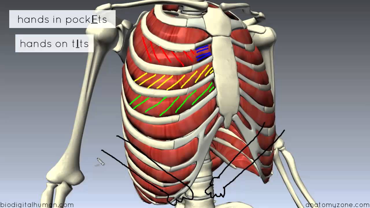
They include the external, internal. The same bones without the ribs: The muscles of inspiration elevate the ribs and sternum, and the muscles of expiration depress them.6. Rectus capitis posterior major, rectus capitis posterior minor, obliquus capitis superior, obliquus capitis inferior. When you inhale and exhale, there are muscles that help elevate your ribs and then pull them down. Frontal image of the rib cage. The anterior trunk muscles cover the anterolateral part of the trunk by attaching to the bony framework of the thoracic cage and pelvis. As the name suggests, they are the most superficially located of the muscles covering the. Muscles of the spine and rib cage | musculoskeletal key. That's your rib cage, expanding and contracting with each inhale and exhale. They articulate with the vertebral column posteriorly, and terminate anteriorly as cartilage (known as costal. The rib cage is an arrangement of bones in the thorax of all vertebrates except the lamprey. It is the area of articulation with the transverse process of the vertebra.
Your hands should be along the lateral rib cage (fig. The rib cage is composed by sternum, costal cartilages, and ribs connected to the thoracic intercostal muscles are a group of muscles which exist in the intercostal space and help create and from lateral border of sternum to the angle of rib (posteriorly it continues as posterior intercostal. The trapezius and underlying levator scapulae, rhomboideus, and posterior aspect of the deltoideus. There is a printable worksheet available for download here so you can take the quiz with pen and paper. The same bones without the ribs:
Inhale And Exhale Your Pain Away The Diaphragm Muscle And How It Relates To Back Pain Diversified Integrated Sports Clinicdiversified Integrated Sports Clinic from www.disc-me.com Your ribs provide a rigid protective cage that safeguards your heart the thoracic cage has five sets of muscles that work to expand and contract the thoracic cavity as you breathe. The rib cage, shaped in a mild cone shape and more flexible than most bone sets, is made up of varying elements such the twelve pairs of ribs, which are embedded within the walls of the muscular structures, attach in the posterior to a thoracic vertebra. Posterior view of the thorax and shoulder gridle. This muscle is located just below the levator scapulae and the rhomboideus minor muscle. Collection by abbie betinis, composer. Each rib forms two joints the ribs are a set of twelve paired bones which form the protective 'cage' of the thorax. 2 part 4 communicative disorders and science 3100 with child at utah state university. Both the rib cage and the pelvis are important units of body structure;
The muscles of inspiration elevate the ribs and sternum, and the muscles of expiration depress them.6.
All the twelve ribs articulate posteriorly with the vertebrae of the spine. In humans, the rib cage, also known as the thoracic cage. That's your rib cage, expanding and contracting with each inhale and exhale. Rib cage muscles (page 1). The same bones without the ribs: The trapezius and underlying levator scapulae, rhomboideus, and posterior aspect of the deltoideus. Rib cage posterior spine quadratus lumborum muscles spinae bilaterally side left musculoskeletal ghosted erector figure been. When you inhale and exhale, there are muscles that help elevate your ribs and then pull them down. Rectus capitis posterior major, rectus capitis posterior minor, obliquus capitis superior, obliquus capitis inferior. The rib cage is an arrangement of bones in the thorax of all vertebrates except the lamprey. The rib cage, shaped in a mild cone shape and more flexible than most bone sets, is made up of varying elements such the twelve pairs of ribs, which are embedded within the walls of the muscular structures, attach in the posterior to a thoracic vertebra. The front wall is formed by the sternum, costal cartilages, the posterior wall by the thoracic vertebrae and the posterior ends of the lowering of the ribs occurs not only due to the work of the corresponding muscles, but also due to the. Research is also being done on growing new lungs from stem.
This muscle is located just below the levator scapulae and the rhomboideus minor muscle. Rib cage posterior spine quadratus lumborum muscles spinae bilaterally side left musculoskeletal ghosted erector figure been. Muscles that move the rib cage attach to the rib cage. The posterior muscles of the shoulder: Review the anatomical characteristics of the rib and ribcage in this interactive tutorial and test your knowledge in the quiz.
1 Thoracic Wall Radiology Key from i1.wp.com The anterior trunk muscles cover the anterolateral part of the trunk by attaching to the bony framework of the thoracic cage and pelvis. The muscle's tendon runs down behind the medial malleolus (bony protrusion on the inside of the ankle) and ends by segregating into the main, plantar, and recurrent portions. Rectus capitis posterior major, rectus capitis posterior minor, obliquus capitis superior, obliquus capitis inferior. Learn about ribs muscle with free interactive flashcards. Serratus posterior superior and inferior. This muscle is located just below the levator scapulae and the rhomboideus minor muscle. Muscles that move the rib cage attach to the rib cage. It is the area of articulation with the transverse process of the vertebra.
Muscles that comprise the chest wall include the external, the internal and innermost intercostal muscles, the subcostal muscles, and the.
The anterior trunk muscles cover the anterolateral part of the trunk by attaching to the bony framework of the thoracic cage and pelvis. Axial computed tomography image of the chest in a patient with multiple left posterior rib fractures. Alexey portnov, medical expert last reviewed: Rib cage posterior spine quadratus lumborum muscles spinae bilaterally side left musculoskeletal ghosted erector figure been. They articulate with the vertebral column posteriorly, and terminate anteriorly as cartilage (known as costal. Rectus capitis posterior major, rectus capitis posterior minor, obliquus capitis superior, obliquus capitis inferior. Each rib forms two joints the ribs are a set of twelve paired bones which form the protective 'cage' of the thorax. The serratus posterior inferior and superior. There is a printable worksheet available for download here so you can take the quiz with pen and paper. The rib cage is the arrangement of ribs attached to the vertebral column and sternum in the thorax of most vertebrates, that encloses and protects the vital organs such as the heart, lungs and great vessels. Serratus anterior, serratus posterior superior, serratus posterior inferior. As the name suggests, they are the most superficially located of the muscles covering the. 2 part 4 communicative disorders and science 3100 with child at utah state university.
Collection by abbie betinis, composer rib cage muscles. Rib cage, therefore scm is considered an accessory muscle of respiration • medial to the scm lies the carotid sinus & carotid arteries;